genome resources
The Animal
(Contributed by Peter Ladurner and Eugene Berezikov)
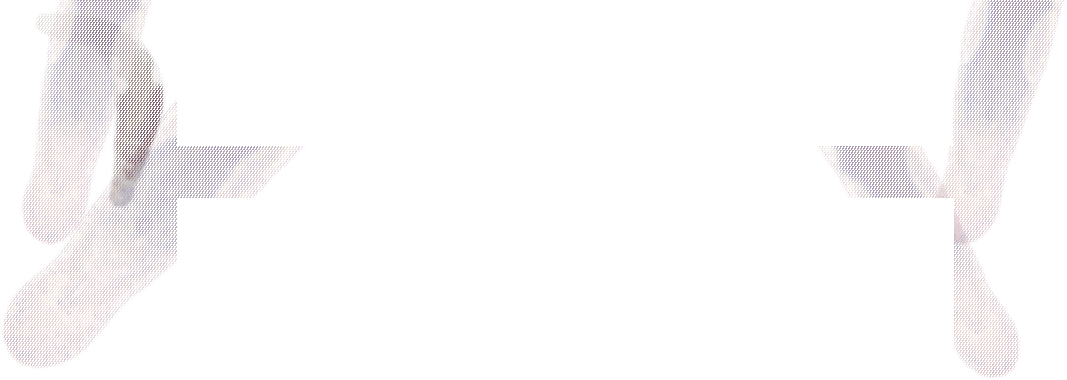
Macrostomum lignano (Fig. 1) is a small (1.5 mm) free-living, marine flatworm which represents a basal member of the Rhabditophora, the largest taxon within the phylum Platyhelminthes. M. lignano is used as a model organism for addressing fundamental questions of flatworm stem cell biology, regeneration, and aging. It is also extensively used to study sex allocation, the question about how simultaneous hermaphrodites distribute energy resources to the male and female reproductive function.
Figure 1. Interference contrast photomicrograph of a living Macrostomum lignano and corresponding schematic drawing. The animal was relaxed and is slightly squeezed. The gut is filled with brownish diatoms, its food item. [Picture out "From Hydra to man" Ladurner et al., 2008]
Natural habitat
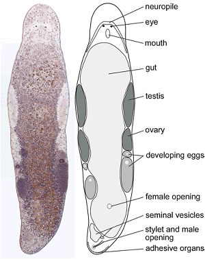
The natural environment of M. lignano is the high-tide interstitial sand fauna of beaches of the Northern Adriatic Sea (Fig. 2).
Figure 2. The natural environment of M. lignano
Culturing in laboratory
M. lignano is easily maintained in the laboratory.
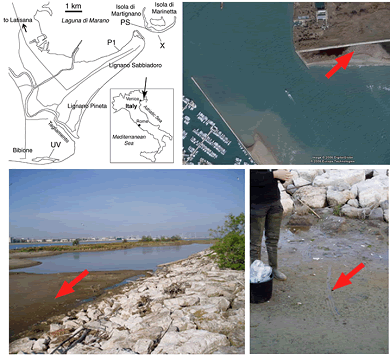
In lab cultures (Fig. 3, Movie 1) M. lignano feeds exclusively on the diatom Nitzschia curvilineata.
Figure 3.Culture of M. lignano in Petri dishes (top left) with worms (white) and diatomes (brownish); (botton left) Box with petri dishes; (top right) climate chamber with boxes on shelfs; (bottom right) diatom on agar cultures in tubes.
Movie 1. M. lignano feeding on diatoms.
Movie 2. Negative phototaxis in M. lignano.
Reproduction
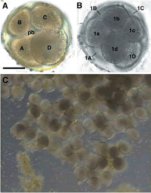
M. lignano is a non-self-fertilizing hermaphrodite. Animals lay single cell eggs of about 100 μm in diameter. Embryonic development (Fig. 4) takes about 5 days until eggs hatch from the eggshell. The generation time is about 18 days.
Figure 4. Embryonic development: Four (A) and 8-cell (B) stage and eggs (C).
Morphology
A multiciliated epidermis completely covers the animal (Fig.5). The cilia cells are responsible for locomotion and the reason why Macrostomum can only swim ahead but not reverse. A complex muscle system (Fig. 6) consisting of circular, diagonal, and longitudinal muscle cells is present below the epidermis. The nervous system consists of a brain with a neuropile, two main lateral- and a dorsal and ventral nerve cord. Two pigment-cup eyes are present for light perception.
In the parenchyma - the tissue between the body wall and the gut - various gland cell types and the stem cells are present. The gut lacks an anus and is completely lined with ciliated gastrodermal cells. In well-fed animals the gut cavity is densely filled with diatoms, which results in a brownish appearance of the animals. Protonephridia are responsible for osmoregulation.
The reproductive system of hermaphroditic animals consists of paired testes and ovaries located in the central region of the animal. Due to the transparency the detailed morphology of structures such as e.g. the testis (Fig. 7) can be observed in live animals. Developing eggs can often be observed in the posterior region of the animals. Sperm are accumulated in the seminal vesicles and transferred with the stylet to the copulation partner. Adhesive organs on the tail plate allow the animal to rapidly adhere and detach from the surface.
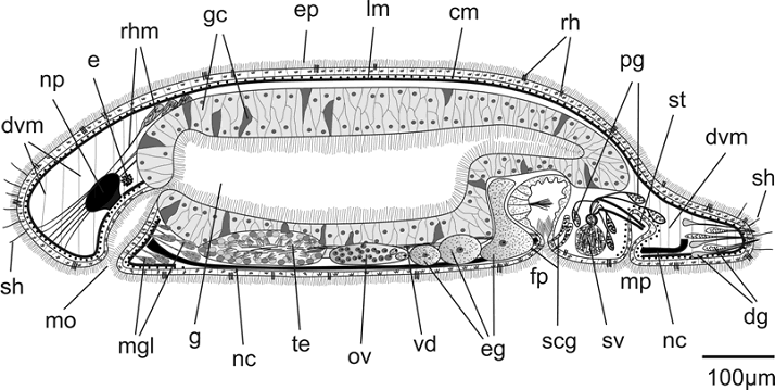
Figure 5. Schematic drawing of Macrostomum sp. midsagittal section. cm - circular muscles, dg - dual gland adhesive system, dvm - dorsoventral muscles, e - eye, eg - eggs, ep - epidermis, fa - female antrum, fp - female genital pore, g - gut, gc - gastrodermal cells, lm - longitudinal muscles, mgl - mouth gland cells, mo - mouth opening, mp - male genital pore, nc - nerve cord, np - neuropile, ov - ovary, pg - prostate glands, rh - rhabdites, rhm - rhamites, scg - shell and cement glands, sh - sensory hairs, st - stylet, sv - seminal vesicle, te - testis, vd - vas deferens.
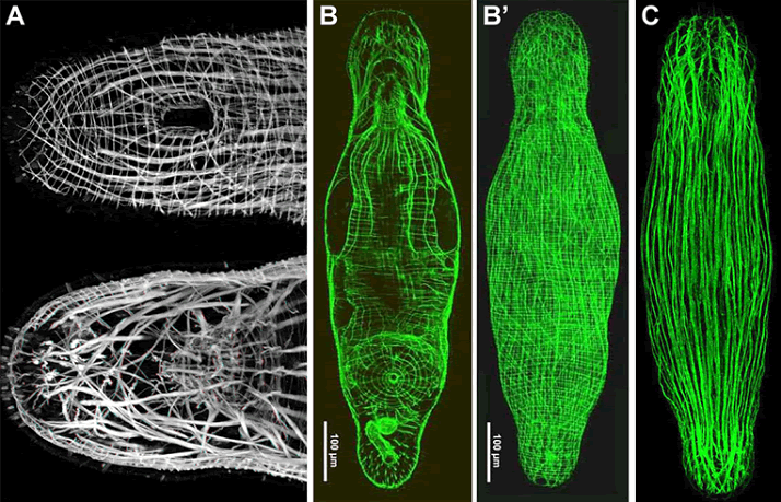
Figure 6. Confocal Projection of phallodin labelled ventral (top) and medio-dorsal muscle system of the head of M. lignano (3D:bottom picture can be viewed with red-green glasses). (B) MMu-1 monoclonal antibody staining of inner muscles (B) and dorsal muscle (B'). (C) MMu-3 monoclonal antibody staining.
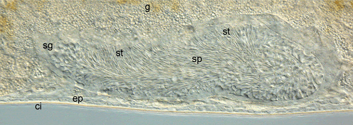
Figure 7. DIC image of testis of a live animal with spermatogonia (sg), spermatids (st) and mature sperm (sp). ep - epidermis; g - gut; ci - bad of beating cilia.
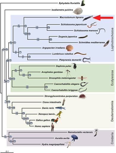
Figure 8.Phylogenetic position of Macrostomum.